Ok so we will skip to my daughter for a minute and let you know what we found out about her. She was born with a VSD. That is just a heart mummer that is actually quite common. But it was a shock to find out after we left the hospital that she had it. Apparently the smaller the hole in the heart the louder it gets. Hers is really loud. Scared a few doctors because it is so loud. A VSD is a hole in the heart that lets blood pass through it. She is followed by cardiology and thankfully we only have to go in every 3 year now instead of every year! I was so excited to her that from the doctor.
Here is what I found on it. Yes it is a lot but it there is some great information.
VSD MummerA
ventricular septal defect (VSD) is a defect in the ventricular septum, the wall dividing the left and right ventricles of the heart.
The ventricular septum consists of an inferior muscular and superior membranous portion and is extensively innervated with conducting cardiomyocytes. The membranous portion, which is close to the atrioventricular node, is most commonly affected in adults and older children.[1][2]
Congenital VSDs are collectively the most common congenital heart defects.
A VSD can be detected by cardiac auscultation. Classically, a VSD causes a pathognomonic holo- or pansystolic murmur. Auscultation is generally considered sufficient for detecting a significant VSD. The murmur depends on the abnormal flow of blood from the left ventricle, through the VSD, to the right ventricle. If there is not much difference in pressure between the left and right ventricles, then the flow of blood through the VSD will not be very great and the VSD may be silent. This situation occurs a) in the fetus (when the right and left ventricular pressures are essentially equal), b) for a short time after birth (before the right ventricular pressure has decreased), and c) as a late complication of unrepaired VSD. Confirmation of cardiac auscultation can be obtained by non-invasive cardiac ultrasound (echocardiography). To more accurately measure ventricular pressures, cardiac catheterization, can be performed.
VSDs are the most common congenital cardiac anomalies. They are found in 30-60% of all newborns with a congenital heart defect, or about 2-6 per 10000 births. It is debatable whether all those defects are true heart defects, or if some of them are normal phenomena, since most of the trabecular VSDs close spontaneously.[4] Prospective studies give a prevalence of 2-5 per 100 births of trabecular VSDs that closes shortly after birth in 80-90% of the cases.
Treatment is either conservative or surgical. Smaller congenital VSDs often close on their own, as the heart grows, and in such cases may be treated conservatively. In cases necessitating surgical intervention, a heart-lung machine is required and a median sternotomy is performed. Percutaneous endovascular procedures are less invasive and can be done on a beating heart, but are only suitable for certain patients. Repair of most VSDs is complicated by the fact that the conducting system of the heart is in the immediate vicinity.
Ventricular septal defect is usually symptomless at birth. It usually manifests a few weeks after birth.
During ventricular contraction, or systole, some of the blood from the left ventricle leaks into the right ventricle, passes through the lungs and reenters the left ventricle via the pulmonary veins and left atrium. This has two net effects. First, the circuitous refluxing of blood causes volume overload on the left ventricle. Second, because the left ventricle normally has a much higher systolic pressure (~120 mm Hg) than the right ventricle (~20 mm Hg), the leakage of blood into the right ventricle therefore elevates right ventricular pressure and volume, causing pulmonary hypertension with its associated symptoms. This effect is more noticeable in patients with larger defects, who may present with breathlessness, poor feeding and failure to thrive in infancy. Patients with smaller defects may be asymptomatic.
Ventricular septal defect (VSD) is the second most common cardiac malformation, accounting for approximately one fifth of all congenital cardiac anomalies. It is usually diagnosed during childhood. In adults, it is diagnosed less often, owing to the fact that, during the patient's early years, large VSDs are corrected surgically, and smaller VSDs close spontaneously.
A VSD is a defect in the interventricular septum, which is composed of muscular and membranous segments. VSDs are classified into 3 main categories according to their location and the appearance of the margins of defects. The clinical significance of the VSD depends on its size and location, the level of pulmonary pressure, and the left ventricular (LV) outflow resistance associated with the VSD. A restrictive VSD produces a small shunt and does not cause significant hemodynamic derangement. In contrast, a large VSD may progressively lead to higher pulmonary resistance and, finally, to irreversible pulmonary vascular changes, producing the so-called Eisenmenger syndrome (reversal of shunt to right-to-left shunt).
Clinically, VSDs produce a characteristic systolic murmur and are associated with recurrent upper respiratory infections. The anatomic localization of all VSDs is facilitated by using 2-dimensional (2D) echocardiographic images with a Doppler system and by superimposing a color-coded direction and velocity of blood flow on the real-time images. Clinically significant VSDs require surgical correction; clinical outcomes are usually excellent.
PathophysiologyThe functional disturbance caused by a ventricular septal defect depends primarily on its size and the status of the pulmonary vascular bed rather than on the location of the defect.
The physical size of the VSD is a major, but not the only, determinant of the size of the left-to-right shunt. The magnitude of the shunt is also determined by the level of pulmonary vascular resistance relative to the systemic vascular resistance. The magnitude of intracardiac shunts is usually described by the Qp:Qs ratio, where Qp is the pulmonary resistance and Qs is the systemic resistance. If the left-to-right shunt is small (Qp:Qs < 1.75:1), the cardiac chambers are not appreciably enlarged, and the pulmonary vascular bed is likely normal. If the shunt is large (Qp:Qs > 2:1), left atrial and LV volume overload occurs, as does right ventricular and pulmonary arterial hypertension. The main pulmonary artery, left atrium, and LV are enlarged.
When a small communication is present (usually <0.5 cm2), the VSD is called restrictive, and the right ventricular pressure is normal. Such a VSD produces a significant pressure gradient between the LV and the right ventricle. It is accompanied by a small (<1.5/1.0) shunt, and it does not cause significant hemodynamic derangement. A small VSD with high resistance to flow permits only a small left-to-right shunt.
A moderately restrictive VSD is accompanied by a moderate shunt (Qp:Qs = 1.5-2.5:1.0) and poses a hemodynamic burden on the LV. This leads to left atrial and LV dilation and dysfunction, as well as a variable increase in pulmonary vascular resistance. Important atrial arrhythmias and, less often, ventricular arrhythmias may occur.
In large nonrestrictive VSDs (usually >1.0 cm2), right and LV pressures are equalized. In these defects, the direction of shunting and the shunt magnitude are determined by the ratio of pulmonary resistance to systemic vascular resistance. Such a VSD results initially in LV volume overload early in life, with a progressive increase in pulmonary artery pressure. The natural history of VSDs has a wide spectrum of findings, ranging from spontaneous closure to congestive cardiac failure and death in early infancy. Within this spectrum are the possible development of pulmonary vascular obstruction, right ventricular outflow tract obstruction, aortic regurgitation, and infective endocarditis.
After patients with a large VSD are born, pulmonary vascular resistance may remain higher than normal, and thus, the size of the left-to-right shunt may initially be limited. As pulmonary vascular resistance continues to decrease in the first few weeks after birth, owing to the normal involution of the media of small pulmonary arterioles, the size of the left-to-right shunt increases. Eventually, a large left-to-right shunt ensues, and clinical symptoms become apparent.
In most cases during early infancy, pulmonary vascular resistance is only slightly elevated, and the major contribution to pulmonary hypertension is the extremely large pulmonary blood flow. However, in some infants with a large VSD, pulmonary arteriolar medial thickness never decreases. With continued exposure of the pulmonary vascular bed to high systolic pressure and high flow, pulmonary vascular obstructive disease develops. When the ratio of pulmonary to systemic resistance approaches 1:1, the shunt becomes bidirectional, signs of heart failure abate, and the patient becomes cyanotic (Eisenmenger physiology).
Frequency
United StatesVentricular septal defects are the second most common congenital anomalies of the heart, accounting for approximately 20% of all congenital cardiac malformations.1, 2
Mortality/Morbidity • Small ventricular septal defects pose an ongoing and relatively high risk of endocarditis.
• Perimembranous or outlet VSDs may be associated with progressive aortic valve regurgitation caused by prolapse of the aortic cusp into the defect.
• The late development of subaortic and subpulmonary stenosis has been reported.
RaceNo particular racial predilection has been reported for ventricular septal defect.
SexNo difference in the incidence of ventricular septal defect in male and female patients has been reported.
AgeVentricular septal defect is usually diagnosed in children.
• Infants: In unusual circumstances, a VSD causes difficulties in the immediate postnatal period, although congestive heart failure during the first 6 months of life is a frequent occurrence. Early diagnosis is helpful in ensuring more careful observation of the affected infant. The examining physician usually suspects the diagnosis because of a harsh systolic murmur at the lower left sternal border. The ECG and chest radiographic findings are within normal limits in the immediate neonatal period because appreciable left-to-right shunting occurs only after pulmonary vascular resistance decreases as the pulmonary vessels lose their fetal characteristics. These infants should be closely monitored.
• Children: After the first year of life, a variable clinical picture emerges in children with VSD. If a small defect is present, the child is usually asymptomatic, the ECG usually appears normal, and the chest radiograph shows normal or mildly increased pulmonary vascular markings. Effort intolerance and fatigue are associated with moderate left-to-right shunts. These children have cardiomegaly with a forceful LV impulse and a prominent systolic thrill along the lower left sternal border. The second heart sound is normally split, with moderate accentuation of the pulmonic component; a third heart sound and rumbling diastolic murmur that reflects increased flow across the mitral valve are audible at the cardiac apex. The characteristic murmur, which results from flow across the defect, is harsh and holosystolic. This murmur is best heard along the third and fourth interspaces to the left of the sternum and is widely transmitted over the precordium. A basal midsystolic ejection murmur caused by an increase in flow across the pulmonic valve also may be heard. The ECG reveals left ventricular hypertrophy or combined ventricular hypertrophy, and the chest radiograph and CT scan show cardiomegaly, left atrial enlargement, and vascular engorgement.
AnatomyThe ventricular septum comprises 4 compartments: the membranous septum; the inlet septum; the trabecular septum; and the outlet, or infundibular, septum.
Defects result from a deficiency of growth or a failure of alignment or fusion of component parts. Defects are most commonly classified as occurring in or adjacent to 1 or more of the septal components.
The most common defects occur in the region of the membranous septum and are referred to as paramembranous or perimembranous defects because they are larger than the membranous septum itself and are associated with a muscular defect at a portion of their perimeter. They also are known as infracristal, subaortic, or conoventricular defects. These perimembranous defects also may be defined by their adjacent areas as an inlet, trabecular, or outlet defect.
A second type of defect is one with an entirely muscular rim. Such muscular defects also may be defined as inlet, trabecular, central, apical, marginal or Swiss cheese, or outlet types. They vary greatly in size, shape, and number.
A third type of defect occurs when the outlet septum is deficient. This is commonly referred to as supracristal, subpulmonary, outlet, infundibular, or conoseptal. Because the aortic and pulmonary valves are in fibrous continuity, this type of defect also may be referred to as a doubly committed subarterial defect.
A septal deficiency of the site of the atrioventricular (AV) septum characterizes defects called AV septal, AV canal, or inlet septal defects.
The other feature of any defect may be a malalignment of the septal components. Either the inlet septum or the outlet septum may be malaligned. Malalignment of the inlet septum produces either mitral or tricuspid valve override and/or straddle. Malalignment of the outlet septum may be to the right or the left of the trabecular septum. When it is to the left of the trabecular septum, the VSD is characteristic of tetralogy of Fallot; double-outlet ventricle; truncus arteriosus; and, in some cases, transposition of the great arteries.
PresentationThe clinical presentation of patients with a ventricular septal defect varies according to the size of the defect and the pulmonary blood flow and pressure.3, 4, 5, 6, 7
SymptomsSmall VSDs with trivial left-to-right shunts and normal pulmonary arterial pressures are the most common. Patients with these VSDs are asymptomatic; the cardiac lesion is usually found during routine physical examination.
Patients with a moderately restrictive VSD often present with dyspnea in adult life. In patients with large nonrestrictive VSDs, the condition frequently progresses to Eisenmenger syndrome.
SignsPhysical examination typically reveals a displaced cardiac apex with a similar holosystolic murmur, as well as an apical diastolic rumble and third heart sound (S3) at the apex, caused by the increased flow through the mitral valve.
Characteristically, a loud, harsh, or blowing holosystolic murmur is best heard over the lower left sternal border in the third or fourth intercostal space. This is frequently accompanied by a thrill. Sometimes, the murmur ends before the second heart sound, presumably because of closure of the defect during late systole.
A short, harsh systolic murmur localized at the apex in a neonate is often a sign of a tiny muscular VSD. In the immediate neonatal period, the left-to-right shunt may be minimal because of higher right-sided pressures; thus, the systolic murmur may not be audible during the first few days of life. In premature infants, the murmur may be heard early because pulmonary vascular resistance decreases more rapidly.
Large VSDs with excessive pulmonary blood flow and pulmonary hypertension are responsible for dyspnea, feeding difficulties, poor growth, profuse perspiration, recurrent pulmonary infections, and cardiac failure in early infancy. Cyanosis is usually absent, but duskiness is sometimes noted during periods of infection or when crying. Prominence of the left precordium is common, as are a palpable parasternal lift, a laterally displaced apical impulse and apical thrust, and a systolic thrill.
The holosystolic murmur of a large VSD is usually less harsh than that of a small VSD and is more blowing in nature because of the absence of a significant pressure gradient across the defect. It is even less likely to be audible in newborns. The pulmonic component of the second heart sound may be increased, indicating pulmonary hypertension. The presence of a mid-diastolic, low-pitched rumble at the apex is caused by increased blood flow across the mitral valve and indicates a Qp:Qs of 2:1 or greater.
Natural historyThe natural course of a VSD depends to a large degree on the size of the defect.
A significant number (30-50%) of small defects close spontaneously, most frequently during the first 2 years of life. Small muscular VSDs are more likely to close (as many as 80%) than membranous VSDs (as many as 35%). The vast majority of defects that close do so before the patient is 4 years of age, although spontaneous closure has been reported in adults.
Most children with small defects remain asymptomatic, without evidence of an increase in heart size, pulmonary arterial pressure, or resistance. One of the long-term risks for these patients is infective endocarditis. Some long-term studies of adults with small VSDs that are not treated with surgery indicate an increase in the incidence of arrhythmia, subaortic stenosis, and exercise intolerance. The Council on Cardiovascular Disease in the Young of the American Heart Association states that an isolated, small, hemodynamically insignificant VSD is not an indication for surgery.
More commonly, infants with large defects have repeated episodes of respiratory infection and heart failure despite optimal medical management. Many of these infants experience heart failure, manifested as a failure to thrive. Pulmonary hypertension occurs as a result of high pulmonary blood flow. These patients are at risk for the eventual development of pulmonary vascular disease if the defect is not repaired.
Patients with VSD are also at risk for the development of aortic valve regurgitation; the risk is greatest for patients with supracristal VSD.
A VSD that either decreases in size or closes completely during the first year of life presents no problem to the practicing physician.
Spontaneous closure occurs by 3 years of age in about 45% of patients born with VSD. In some patients, spontaneous closure does not occur until 8-10 years or age, or even later. Spontaneous closure is more common in patients born with a small VSD; nonetheless, about 7% of infants with a large defect who experience congestive heart failure early in life also experience spontaneous closure. Partial, rather than complete, closure is common in patients with both large and small VSDs. Spontaneous closure of a perimembranous VSD (from tricuspid leaflet tissue apposition) or of a small muscular VSD during adulthood is uncommon (<10%).
Preferred Examination
Echocardiography Two-dimensional and Doppler color-flow mapping may be used to identify the type of defect in the ventricular septum. Perimembranous VSDs are characterized by septal dropout in the area adjacent to the septal leaflet of the tricuspid valve and below the right border of the aortic annulus.
The subaortic or anterior malalignment type of VSD appears just below the posterior semilunar valve cusps, entirely superior to the tricuspid valve. Subpulmonary VSD appears as echo dropout within the outflow septum and extending to the pulmonary annulus. One or two of the aortic cusps may be seen to be protruding through the defect into the right ventricular outflow tract. The inlet AV septal-type of VSD extends from the fibrous annulus of the tricuspid valve into the muscular septum; it is often entirely beneath the septal tricuspid leaflet.
Muscular defects may appear anywhere throughout the ventricular septum. They may be either large and single or small and multiple.
The anatomic localization of all VSDs is facilitated by coupling 2D sonograms with a Doppler system and by superimposing a color-coded direction and velocity of blood flow on the real-time images.
Chest radiographyIn patients with small VSDs, the results of chest radiographs are usually normal. With medium-size VSDs, minimal cardiomegaly and a borderline increase in pulmonary vasculature may be observed. In large VSDs, the chest radiograph shows gross cardiomegaly with prominence of both ventricles, the left atrium, and the pulmonary artery. The pulmonary vascular markings are increased, and frank pulmonary edema, including pleural effusions, may be present.
ElectrocardiographyThe ECG mirrors the size of the shunt and the degree of pulmonary hypertension.
Small restrictive VSDs usually produce a normal tracing. Medium-size VSDs produce a broad, notched P wave characteristic of left atrial overload. Signs of LV volume overload — namely, deep Q and tall R waves with tall T waves in leads V5 and V6 — are present. In addition, signs of atrial fibrillation are often present. Large VSDs produce right ventricular hypertrophy with right-axis deviation. With further progression, the ECG shows biventricular hypertrophy; P waves may be notched or peaked.
Limitations of TechniquesThe 2D echocardiogram shows the position and size of the ventricular septal defect. Small defects, especially those of the muscular septum, may be difficult to image; they might be visualized only by means of color Doppler examination.
In defects of the membranous septum, a thin membrane may partially cover the defect and limit the volume of the left-to-right shunt. Although the membrane is called a ventricular septal aneurysm, it consists of tricuspid valve tissue.
I hope this information helped you as much as it helped me understand things better.
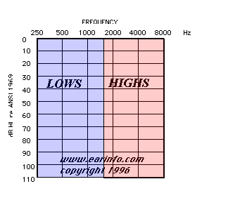
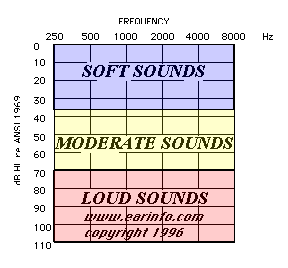
 The Cochlea


